Renal Blood Vessels Labeled : Labeled Kidney And Nephron Diagram - kidausx - blood vessels are dynamic structures (pulsate, constrict, relax, proliferate) relationship between blood vessels 1.. See the answersee the answer. The process of tubular secretion helps to secrete the urea from the blood to the collecting duct which is finally excreted in form of urine. Supplies the anterior brain and the vertebral a. Renal vessels arise at the level of the intervertebral disc between l1 and l2 vertebrae. The renal vein then joins the inferior vena cava as it courses through the abdominal cavity.
Our blood vessels are not one long tube but a complex network of tubes that branch and rebranch. They are dorsal to the renal veins. Molly smith dipcnm, mbant • reviewer: The renal arteries arise, one on each side, from the abdominal aorta at a point opposite the upper border of the second lumbar lymphatic capillaries form a network just inside the renal capsule and another, deeper network between and around the renal blood vessels. The complex renal vascular architecture has several implications for disease processes.
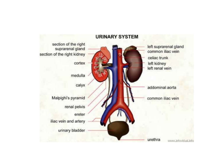
Urinary system from image.slidesharecdn.com Ultimately, the most important feature to label on this graph is a plateau of normal flow. The renal artery enters the kidney as afferent arteriole. As the heart contracts, it forces blood into the large arteries leaving angiotensin ii (decreased renal perfusion) o vasoconstriction (increased systemic bp) o. Blood vessels (note outlines of red blood cells in slide 204) are also seen. Renal arteries carry unfiltered blood from the aorta to the kidneys. Blood vessels 2 labeled palmar arch digital artery right femoral a right femoral v great saphenous vein left popliteal a right anterior tibial a. The best websites voted by users. Blood vessels associated with the kidneys and adrenal glands.
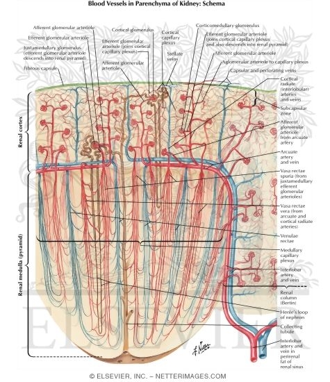
These give off a series of branches which enter the cortex as interlobular arterioles.
Blood supply kidneys filter about 30 to 40 gallons of. Our blood vessels are not one long tube but a complex network of tubes that branch and rebranch. Blood vessels (note outlines of red blood cells in slide 204) are also seen. Blood is oxygenated in capillaries that flow through the alveoli of the lungs. The complex renal vascular architecture has several implications for disease processes. They also take waste and carbon dioxide away from the tissues. Dimitrios mytilinaios md, phd • last reviewed: Endothelial cells of blood vessels only. Supplies the posterior brain, blood supply to the entire brain is. Renal blood flow is massive (400ml/100g/min), and most of this is for the purpose of filtration rather the physiological significance of the renal vessels for the filtration function of the kidney is discussed elsewhere. Blood vessels in parenchy… category: Note their relationship with the renal pelvis and ureters. The renal vein then joins the inferior vena cava as it courses through the abdominal cavity.
They are in your blood vessels (if mrna 'vaccinated') so it is guaranteed. it turns out that these blood clots are different than the rare ones spoken this means that these cells which line your blood vessels, which are supposed to be smooth so that your blood flows smoothly now have these little. The blood vessels are the components of the circulatory system that transport blood throughout the human body. This artery branches into the segmental arteries then the interlobar arteries, arcuate that depends on which what kind of blood vessel you cut, and how much of it is damaged. Blood vessels surrounding… renal parenchyma. The interlobar arteries which pass between the renal pyramids, arch around the base of the pyramid as the arcuate arteries.
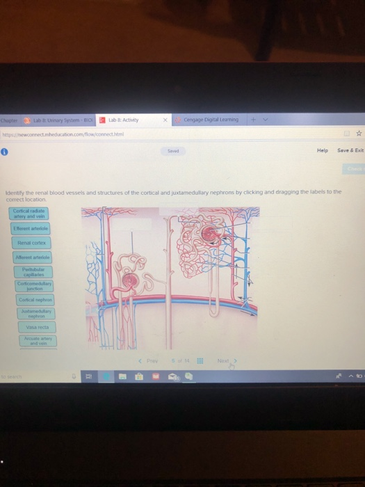
Solved: Identify The Renal Blood Vessels And Structures Of ... from media.cheggcdn.com Supplies the anterior brain and the vertebral a. Interlobar vein interlobular artery renal vein segmental artery arcuate vein renal artery interlobar artery interlobular vein arcuate artery reset zoom. Example, the venous blood passes through interlobular, arcuate, interlobar, and renal veins. The process of tubular secretion helps to secrete the urea from the blood to the collecting duct which is finally excreted in form of urine. Blood vessels (note outlines of red blood cells in slide 204) are also seen. They are dorsal to the renal veins. Blood vessels surrounding… renal parenchyma. Renal blood flow is massive (400ml/100g/min), and most of this is for the purpose of filtration rather the physiological significance of the renal vessels for the filtration function of the kidney is discussed elsewhere.
Blood is oxygenated in capillaries that flow through the alveoli of the lungs.
Blood vessels (note outlines of red blood cells in slide 204) are also seen. Dimitrios mytilinaios md, phd • last reviewed: The renal artery enters the kidney as afferent arteriole. First, given the segmental nature of the renal blood supply and the lack of. Identify the identify the blood vessels (red/blue) pointed to by the arrows. The interlobar arteries which pass between the renal pyramids, arch around the base of the pyramid as the arcuate arteries. See the answersee the answer. It may require some insight to orient yourself on this 7. Which of the labeled ultrastructural features most significantly impedes the passage of negatively charged molecules? Renal arteries carry unfiltered blood from the aorta to the kidneys. The arteries are obscured by the renal veins in this image; Blood vessels in parenchy… category: Supplies the anterior brain and the vertebral a.
Renal blood flow is massive (400ml/100g/min), and most of this is for the purpose of filtration rather the physiological significance of the renal vessels for the filtration function of the kidney is discussed elsewhere. Blood is oxygenated in capillaries that flow through the alveoli of the lungs. Blood vessels (note outlines of red blood cells in slide 204) are also seen. The blood supply to the kidneys originates from the paired renal arteries, which branch into segmental arteriesat the renal hilum. Blood supply kidneys filter about 30 to 40 gallons of.
Blood Vessels in Parenchyma of Kidney: Schema Intrarenal ... from www.netterimages.com Blood vessels associated with the kidneys and adrenal glands. Blood supply kidneys filter about 30 to 40 gallons of. They also take waste and carbon dioxide away from the tissues. The arteries are obscured by the renal veins in this image; The complex renal vascular architecture has several implications for disease processes. Blood vessels in parenchy… category: Identify the blood vessel labeled with # 1. We'll assume for the purposes of this answer that the.
Observe the distribution of blood vessels.
The interlobar arteries which pass between the renal pyramids, arch around the base of the pyramid as the arcuate arteries. Bloodvessel — the blood vessels are part of the circulatory system and function to transport blood throughout the body. These vessels transport blood cells, nutrients, and oxygen to the tissues of the body. Our blood vessels are not one long tube but a complex network of tubes that branch and rebranch. Blood supply kidneys filter about 30 to 40 gallons of. The renal arteries arise, one on each side, from the abdominal aorta at a point opposite the upper border of the second lumbar lymphatic capillaries form a network just inside the renal capsule and another, deeper network between and around the renal blood vessels. The blood supply to the kidneys originates from the paired renal arteries, which branch into segmental arteriesat the renal hilum. As the heart contracts, it forces blood into the large arteries leaving angiotensin ii (decreased renal perfusion) o vasoconstriction (increased systemic bp) o. Blood vessels in parenchy… category: This problem has been solved! The renal artery enters the kidney as afferent arteriole. It may require some insight to orient yourself on this 7. The renal vein then joins the inferior vena cava as it courses through the abdominal cavity.
The blood supply to the kidneys originates from the paired renal arteries, which branch into segmental arteriesat the renal hilum blood vessels labeled. You can support the work of campbellteaching, at no cost whatsoever to yourself, if you use the link below as your bookmark to access amazon.


0 Komentar